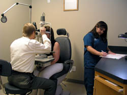
Testing & Eye Exams
 Regular
glaucoma check-ups are the most effective method to prevent sight loss due
to the disease. Testing for glaucoma is completely painless, and early detection
is extremely important. Once your vision is impaired by glaucoma, the damage
is permanent and cannot be reversed.
GIA offers a comprehensive array of glaucoma tests to provide an accurate glaucoma diagnosis. These tests include:
- Applanation Tonometry
- Ophthalmoscopy
- Perimetry
- Gonioscopy
- Optical Coherence Tomography
- Fundus Photography
- Pachymetry
Applanation tonometry measures the pressure within your eye. We use eye drops to numb the surface of the cornea, then we use a tool called a tonometer to apply light pressure (applanation) to the eye to measure intraocular pressure.
The range for normal pressure is 12-22 mm Hg (“mm Hg” refers to millimeters of mercury, a scale used to record eye pressure). Most glaucoma cases are diagnosed with pressure exceeding 20 mm Hg. However, some people can have glaucoma at pressures lower than 20 mm Hg. Remember, eye pressure is unique to each person.
This procedure examines your optic nerve for glaucoma damage. First, we use eye drops to dilate the pupil in order to examine the interior of the eye. An ophthalmoscope is used to examine the shape and color of the optic nerve.
A nerve that is cupped or not a healthy pink color is cause for concern. If your intraocular pressure is not within the normal range or if the optic nerve looks unusual, we may perform additional special glaucoma tests: perimetry, gonioscopy or optical coherence tomography.
Perimetry is a visual field that tests your peripheral vision. This test will help to determine whether your vision has been affected by glaucoma. During this test you will be asked to look straight ahead and signal when you see spots of light
You may need to repeat this test on a future visit to compare the results to your initial visual test. If glaucoma has been diagnosed, a visual field test may be done once or twice yearly to check for any changes in your vision.
This procedure helps to determine whether the angle where the iris meets the cornea is open and wide, or narrow and closed. Before this exam, eye drops are used to numb the surface of the cornea
For more precise imaging of the optic nerve, we use optical coherence tomography. This works similarly to an ultrasound, but instead of measuring sound, it measures the reflection of infrared light, which is reflected uniquely by different tissues. This test can detect glaucoma much earlier than other current methods.
Fundus photography is a highly specialized form of medical imaging that takes photos of the back of your eye by focusing light through the cornea, pupil and lens. These pictures are use to assess the health of the optic nerve. The photographs are used for comparison, documentation, and sometimes to diagnose glaucoma.
Recent research has identified a relationship between eye pressure and the thickness of the cornea. Measurement of cornea thickness, called pachymetry, is important when making a diagnosis of glaucoma. It is also important when evaluating intraocular pressure control for patients receiving treatment.
Research has shown that people with thin corneas may have much higher intraocular pressures than previously determined using the standard eye pressure measurement technique called applanation tonometry. Similarly, a thicker than normal cornea may mean that the eye pressure is actually much lower than determined by standard applanation tonometry.

