Microsurgery
 Sometimes
treatment with medication and laser surgery doesn't lower eye pressure suffciently.
In these cases, microsurgery may be required.
Sometimes
treatment with medication and laser surgery doesn't lower eye pressure suffciently.
In these cases, microsurgery may be required.
Trabeculectomy involves creating a tiny drainage hole in the sclera (the white part of the eye) that allows fluid to flow out of the eye, lowering eye pressure. This prevents or reduces damage to the optic nerve.
The surgery is usually done with either a local anesthetic or a limited type
of anesthesia called intravenous sedation. In addition, an injection is given
around or behind the eye to prevent eye movement. You will be relaxed and
drowsy, and will not experience any pain during surgery.
After surgery, the eye will be red and irritated shortly after surgery and
there may be increased eye tearing or watering. The inner eye fluid flows
through the surgically-created hole and forms a small blister-like bump called
a bleb. The bleb, usually located on the upper surface of the eye, is covered
by the eyelid, and is usually not visible.
Occasionally, the surgically-created drainage hole begins to close and the pressure rises again. This happens because the body tries to heal the new opening in the eye, as if the opening were an injury. This rapid healing occurs most often in younger people, because they have a stronger healing system. Anti-wound healing drugs, such as mitomycin-C and 5-FU, help slow down the healing of the opening. If needed, glaucoma filtering surgery can be done a number of times in the same eye
Another type of microsurgery involves the placement of glaucoma drainage devices (also known as setons, valves, tubes or shunts). It is usually recommended when the eye pressure cannot be lowered enough with medicine, laser treatment, or trabeculectomy, and also for certain specific types of glaucoma.
Valve implantation is performed in an operating room on an outpatient basis. A local anesthetic, which is given by injection at the side of the eye, is used to prevent discomfort during the operation. Sedation will be given before the anesthetic to relax you.
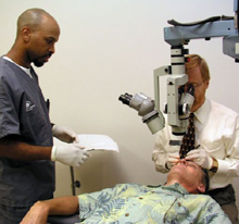
The surgery requires a special microscope, which is suspended over your eye. This enables your doctor to see clearly the very fine details of the eye. Tiny stitches, thinner than a human hair, are used to close the surgical incision. This surgery is performed in about an hour. It may take longer if there has been previous eye surgery, inflammation, abnormal vessels, or eye problems.
Recovering from microsurgery
Immediately following glaucoma surgery, the eye is soft and delicate. Your physical activity should be restricted to avoid lifting, bending, and straining. Vision may be quite blurred during the recovery period. Your care after the surgery is as important to the long-term success of the operation as the surgery itself.
Each person heals differently after surgery. Therefore, your care must be individually adjusted depending upon the appearance of the tissues and the condition of the eye.
Patients are typically seen many times during the first 6 weeks following surgery so your doctor may observe your eye and adjust your treatment. As the eye heals, office visits become less frequent.
*Supplemental information courtesy of
Glaucoma Research Foundation

